
![]()
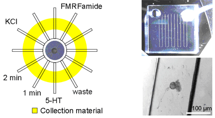
The response of a neuron to chemical or electrical stimulation depends on the chemical environment surrounding the cell. In life, different regions of a neuron experience distinct chemical environments. Microfluidic devices provide a suitable experimental platform to study the peptide release from stimulated neurons; they enable maximal control of the microenvironments surrounding different subregions of individual neurons and manipulation of mass-limited samples. For this purpose, we have developed several microfluidic chip designs that offer a controlled environment for culturing a single or small number of neurons and collecting peptide releasates depending on chemical stimuli with spatial and temporal resolution using octadecyltrichlorosilane (C18) modified surfaces or porous polymer monolith fabricated in a fused silica capillaries. The collected peptide releasates are characterized by matrix-assisted laser desorption/ionization (MALDI) mass spectrometry (MS) as well as MALDI MS imaging techniques, which provide insights on neuron microcircuit formation and damage repair. This work is performed in collaboration with Gillette and Nuzzo. |
Schematic diagram and images of microfluidic devices |
| Relevant Publication: K. Jo, M.L. Heien, L.B. Thompson, M. Zhong, R.G. Nuzzo, J.V. Sweedler, Mass spectrometric imaging of peptide release from neuronal cells within microfluidic devices, Lab Chip 7, 2007, 1454-1460. |
 .
.